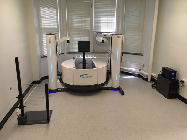
The Orthopedic Functional Imaging Research Laboratory (OFIRL) was founded in 2020 by collaborations between orthopedic surgeons and the University of Iowa Orthopedic Biomechanics Laboratory. Its goal is to advance the understanding of orthopedic care through functional imaging and analysis techniques like weight bearing CT, pedobarography, gait analysis, and more, with focus on Foot and Ankle Surgery. Our lab partnered with CurveBeam to obtain the first 3D weight bearing images of the entire lower extremity (including the hip) in the world using the CurveBeam HiRise System. This low dose weight bearing CT scanner has enabled us to take on projects evaluating combined deformities of the entire lower extremity, as well as isolated foot and ankle pathologies such as progressive collapsing foot deformity (PCFD, aka “flatfoot”), cavovarus deformities, Charcot-Marie-Tooth foot deformities, and many more. The system also allows for imaging of the upper extremities, such as the hand, wrist, and elbow.
The partnership with Paragon28 has optimized our capabilities to perform automatic segmentations and automatic measurements in the foot and ankle. This has enabled large scale analysis leading to future studies and development.
We look forward to continuing to expand our capabilities and collaborations.